In order for a flow cytometer to perform complex analysis like determining whether your cell is a CD45high/CD3+/CD4+/CD25+ cell which has no CD16, is viable and at a specific state of the cell-cycle (a 7 colour, 9 parameter T-cell assay), we first must make sure that the cytometer can look at cells one at a time. This is critical as two cells going through at the same time (co-incident event) will appear to be positive for several markers which may not be true of the individual cells.
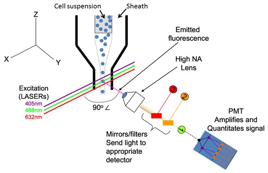
Cytometers establish this single cell stream through hydrodynamic focusing, which is a fancy way of saying focused, non-turbulent (laminar) flow (see Figure 1 below). In this way, the cytometer takes in a “core” sample of cells and uses a surrounding volume of fluid (sheath fluid) to push the whole thing through a flow cell, where all of the fluorescence and light scattering measurements of each cell can be made. This sheath fluid moves through the system and the cell sample is “injected” into the cytometer, focusing everything into a very small (~10μm) stream which promotes a “single-file” line of cells.

Figure 1: Sample is injected into a larger volume of sheath fluid and hydrodynamically focused into a single-cell stream. The point of interrogation where the laser (green) intercepts each cell is where measurements of light scatter and fluorescence are made. Note that the fluid flow (Z axis), laser beam (X axis) and fluorescence collection lens (Y axis) are all at 90º to one another to minimize background and maximize sensitivity.
Subsequently, we must efficiently measure your selected fluorophore(s) and the emitted fluorescence must be matched to the cytometer. The most commonly found lasers on a flow cytometer are 488nm, 405nm and 632/635/640nm which are appropriate for a vast majority of commonly used fluorescent probes. A list of these probes can be found on the flow cytometry room door and on the internet. Each cytometer has a lens just like you would find on a microscope which directs the emitted fluorescence to several photomultiplier tubes (PMTs) which amplify the (typically weak) fluorescence signal (see also Figure 1). In front of each PMT, there is a bandpass filter and/or a shortpass filter or a longpass filter that allows only light of a specified wavelength to reach the detector. It is this wavelength range that we must pay attention to as it dictates which emitted fluorescence is detected by each PMT. For example, a “FITC” channel can be outfitted with a 550/50 bandpass filter which accepts light maximally at 550nm and is 50nm “wide” (i.e. it accepts light from 525-575nm). It is critical to understand the excitation and emission maxima for your intended fluorescent probes and a cheat sheet for this is available here.